Color Vision
Color vision is accomplished in the same manner as black-and-white
imaging, and using the same mechanism. The cyclic nature of the process is identical,
and 11-cis-retinal is the key component, as in the cones.
The difference lies in the protein moieties, not in the retinal.
The scotopsin of the rods is sensitive to all wavelengths of light and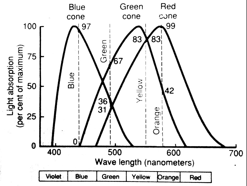 will absorb energy from any part of the visible spectrum, causing
it to dissociate. But the pigments in the cones are wavelength-specific. Some
will respond only to blue, some to red, and some to green wavelengths, and not
to others. This is a matter of their physical structure, which gives them an
absorption peak in a very narrow band of the spectrum. The graph presented here
shows this diagrammatically. The wavelength-specific cones respond to a color
(which is a mixture of various wavelengths) in proportion to their sensitivities.
If the light falling on the cones is a mixture of red and yellow wavelengths,
producing the color orange, the red-sensitive cones are maximally stimulated
by the red component, and the green-sensitive ones less so. The blue-sensitive
ones aren't stimulated at all. The brain sorts this out in the visual cortex
as "orange" based on the proportional response of the different types of cones.
Thus color vision truly resides in the brain. Even animals that are supposedly
"colorblind" have the anatomical apparatus needed for color perception, but
may not process the signals in the CNS to recognize a given mixture in the same
way humans do.
will absorb energy from any part of the visible spectrum, causing
it to dissociate. But the pigments in the cones are wavelength-specific. Some
will respond only to blue, some to red, and some to green wavelengths, and not
to others. This is a matter of their physical structure, which gives them an
absorption peak in a very narrow band of the spectrum. The graph presented here
shows this diagrammatically. The wavelength-specific cones respond to a color
(which is a mixture of various wavelengths) in proportion to their sensitivities.
If the light falling on the cones is a mixture of red and yellow wavelengths,
producing the color orange, the red-sensitive cones are maximally stimulated
by the red component, and the green-sensitive ones less so. The blue-sensitive
ones aren't stimulated at all. The brain sorts this out in the visual cortex
as "orange" based on the proportional response of the different types of cones.
Thus color vision truly resides in the brain. Even animals that are supposedly
"colorblind" have the anatomical apparatus needed for color perception, but
may not process the signals in the CNS to recognize a given mixture in the same
way humans do.
Click Back to return
Or Go To:
Main Page | Corneoscleral Tunic | Uveal Tunic | Retinal Tunic | Physiology of Vision | CNS Processing of Visual Signals